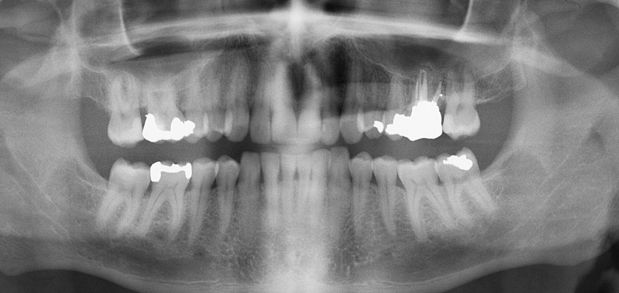
Detect lesions diseases conditions of the teeth 2. - Radiographs dental X-rays are images of your teeth that your dentist uses to assess your oral health.

Types Of Dental Radiographs And Their Uses Dentalnotebook
A radiographic image is formed by a controlled burst of X-ray radiation which penetrates oral structures at different levels depending on varying anatomical densities before striking the.

. Having a cone beam also attracts new patients who expect a highly skilled doctor using the latest techniques. For periodontal diagnosis the high resolution of intraoral radiography helps the visualization of the bony supporting tissues including small details such as periodontal ligament space lamina dura and bony trabecularization3 Digital imaging allows measuring bone loss extent using image analysis tools. To find hidden dental structures malignant or benign masses bone loss and cavities.
Eval growth development 6. This leads to more efficient patient treatment in fewer visits. In the case of 3D dental imaging the advantages are clear granting practitioners and patients alike a better clinical experience.
Illustrate changes secondary to caries periodontal disease trauma 7. CBCT is categorized into large medium and limited volume units based on the size of their field of view FOV. All images can be obtained without the need to reposition the patient.
The patient bites down on the tab so the image will show both top and bottom teeth. Early detection of dental complications save you time the discomfort associated with dental complications and money. Describe the uses of dental imaging.
A 3D scan can help gain a better view of bone structures such as adjacent root positions in order to locate canals and root fractures as well as provide the ability to more. Uses of digital imaging-To detect lesions diseases and conditions of teeth and surrounding structures-Confirm or classify suspected disease-Provide information during dental procedures root canal therapy instrumentation and surgical placement of. It has been used extensively for evaluating dental and osseous disease in the jaws and temporo-mandibular joints and treatment planning for dental implants.
Describe the use of dental imaging. Dentists use bite-wings to get a picture of the back posterior teeth. Dental x-rays can also help your dentist evaluate injuries to your face and mouth.
First because so much data is captured in a single X-ray done right onsite diagnoses are quicker and more accurate. Prior to any extraction x-rays typically need to be taken to identify any possible complicating factors such as whether the roots of the tooth may be running close to a nerve. There are many benefits that come with using high technology systems when getting x-rays and images taken.
Dentists use radiographs for many reasons. Evaluate growth and development. It is safe for patients who are allergic to the contrast agent.
Diagnostic Evaluation of all smile components Measurement of tooth dimensions Pre-Planning desired results Advanced cases Patient Communication Mouth mirror is ineffective Allows them to see what we see Educates with facts Before After Procedure pages Imaged full-face smiles Yourtreatment capabilities Laboratory Communication. Document condition of a patient at a specific point and time 8. Bite-wing x-rays are the type that most people are familiar with.
We have listed 4 of the benefits below to using Advanced Dental Imaging. Digital dental radiographs allow your dentist to examine areas in your mouth that are not visible to the human eye thus facilitating the detection of oral complications in their early stages. - Low-level radiation X-rays are used to acquire images of the interior of your teeth and gums.
Locate abnormalities in surrounding hard and soft tissues. Dental radiographs help dentists assess the development of teeth and bones in children. Uses of Dental Photography.
Willis July 21 2017 Short- Answer Questions 1. Detect dental caries in the early stages. They are not typically done on front anterior teeth.
It is capable of seeing inflammatory processes. It allows dentists to get a fuller picture of the anatomical structure of the patient. Dental x-rays help your dentist identify diseases and problems.
Uses of Dental Images are. X-rays are used to evaluate the amount of available bone tissue and check for other factors that will determine if the patient is a good candidate for dental implants. Provide info during dental procedures 5.
The advantages of digital radiography and the uses in oral implantology are. Confirm or classify suspected disease 3. Dental clinics continue to use 3D dental imaging systems for a variety of clinical applications such as dental implant planning visualizing the progress of abnormal teeth diagnosing root canal issues diagnosing dental cavities or diagnosing dental trauma.
Panoramic X-rays are used to plan treatment for dental implants detect impacted wisdom teeth and jaw problems and diagnose bony tumors and cysts. Periapical and cephalometric radiographs have. A 3D unit also allows general dentistry practices to.
A dental 3D scan allows clinicians to view dental anatomy from different angles. Identify bone loss in the early stages. Better Accuracy Everyone wants good teeth and that means when you get an.
Provide information during dental procedures such as root canal therapy Document a patients condition at a specific time. The history of dental imaging began in the late 1800s with the development of the x-ray image. Localize lesions or foreign objects 4.
Katherine Moran Radiology Mrs. The uses of dental imaging are checking patients oral health and making it clearer to diagnose certain teeth problems such. They get their name from a tab on the x-ray film.
Panoramic films are used for forensic and legal purposes to identify otherwise unrecognizable bodies after. LA Imaging believes in the power of using Advanced Dental Imaging. Dental radiographs are commonly called X-rays.
In 1973 computed tomography CT created images by combining x-ray and computer technology to capture thin slices of tissue1 After that magnetic resonance imaging MRI allowed soft tissue analysis. One of the most significant recent advances in dental radiology is the advent of digital technology which has allowed reduction of numerous limitations of conventional intraoral radiography. Those mentioned above are just some of the benefits of MRI and CT scans in the dental practice.

History Benefits Of Dental X Ray Imaging Artistic Touch Dentistry

Types Of Dental Radiographs And Their Uses Dentalnotebook

What Is Opg X Ray Information About Uses Diagnosis And Treatment

0 Comments